For Vets:

Share this article
Cataract Surgery for Dogs and Cats
Cataract Surgery for Dogs and Cats

Share this article
Cataract Surgery for Dogs and Cats
Cataract Surgery for Dogs and Cats
Share this article
If you notice a cloudy or milky hue in your pet's eyes, it may be a sign of cataract development. Cataracts are a medical condition that affects both animals and humans, and are a leading cause of blindness in dogs. As time goes by, you may notice signs of worsening vision in your pet, such as bumping into furniture, a loss of confidence in climbing and jumping, and overall clumsiness.
When this happens, cataract surgery may be necessary to help your furry friend restore their eyesight and improve their quality of life.
What Are Cataracts?
Cataracts are cloudy or opaque spots that form within the transparent lens of the eye, causing vision to turn blurry and images to lose clarity. An eye is like a camera. It has a clear lens that focuses light on the back of the eye (retina), producing the images we see. When these spots form, the image is disrupted, causing partial blindness.
As cataracts progress, they can cause eye inflammation, retina damage or glaucoma, which may lead to permanent blindness.
What Is Not a Cataract?
Not every occurrence of cloudy eyes is caused by cataracts. Senile nuclear sclerosis is commonly seen in dogs and cats 7 years and older. Lens fibers are continuously being produced. As older fibers are compressed in the nucleus, the lens takes on a bluish-grey hue. No treatment is required as light can pass through a sclerotic lens. The cloudiness does not impair vision.
What Causes Cataracts?
When cataracts occur in younger dogs (less than 6 years of age), it is usually hereditary. Meanwhile, the most common cause of cataracts in cats is uveitis, an inflammation of the eye caused by viral infections.
Diabetes is another leading cause of cataracts in dogs. Early detection and surgery is vital, as these cataracts can progress rapidly. Studies show that 75% of dogs with diabetes will develop cataracts within the first year of diagnosis, while diabetic cats are slightly more resistant to developing cataracts.
Apart from diabetes and uveitis, cataracts can also develop from:
- Old age
- Trauma or injuries to the eye
- Nutritional deficiency
- Toxins
If your pet’s eyes look cloudy or bluish-grey, schedule an examination by a veterinary ophthalmologist. A series of diagnostic tests will be performed (e.g., to check tear production, measure intraocular pressure (IOP), evaluate ocular surface with fluorescein stain test). Advanced testing may include electroretinography (ERG) to evaluate the electrical activity of the retina. If the retina is not functioning properly, cataract surgery will not visually benefit your pet.
Cataract Surgery
Surgical Procedure
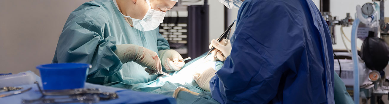
Cataract surgery is performed under general anaesthesia. A nerve blocker will be administered to position the eye correctly. A small corneal incision is made to gain entry into the eye and a blue dye is injected to stain the lens and make it clearly visible for the surgery.
A small window is created in the lens capsule, allowing the surgeon to remove the cataract. An artificial lens implant (intraocular lens) is then inserted into the lens capsule. This replacement lens allows light to focus clearly onto the retina again.
After the corneal incision is closed up with absorbable sutures, the patient is monitored closely as it recovers from anaesthesia. Post-operative eye medications are administered, and eye pressure is measured at regular intervals to ensure it is within normal range.
Post-Surgery
Vets will generally recommend that your pet stay a few days for monitoring following cataract surgery. Intensive aftercare will be required after you bring your pet home, including oral medications and eye drops. Follow-up exams must be scheduled to check for any post-surgery complications.
How Do I Care For My Pet After Surgery?
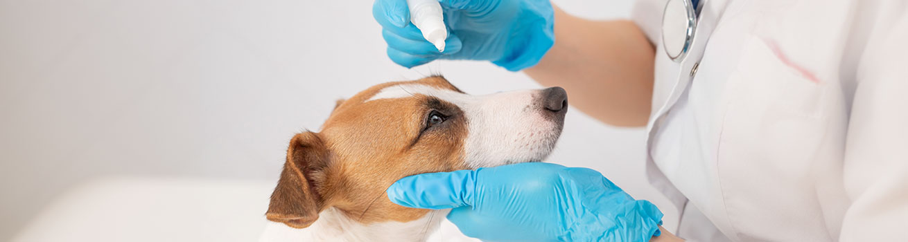
Vision usually improves within 24 hours and continues to improve over several weeks. Your pet will require the following:
- Oral medication
- Medicated eye drops
- E-collar to prevent self-trauma to eyes
- Regular re-examination of the eyes to monitor recovery progress
FAQs
What is the Success Rate of Cataract Surgery?
Cataract surgery for dogs and cats is a common procedure, with a high success rate of 90%. The effectiveness of the surgery may differ depending on the severity of the cataracts.
Early diagnosis and treatment of cataracts increases the success rate and improves long-term outcomes. Scheduling yearly to twice-yearly check-ups for your pets can help identify signs of cataracts before they progress.
What Are the Risks of Cataract Surgery?
The most common postoperative complications are eye inflammation and glaucoma. Other rarer complications include corneal ulcers, infection and retinal detachment.
Does Age Affect Cataract Surgery?
Cataracts can occur in both young and old pets, and age is usually not a consideration in cataract surgery for dogs and cats. However, pets with heavily advanced cataracts or pre-existing medical conditions may not be suited to surgery.
How Quickly Do Pets Recover From Cataract Surgery?
If the surgery is successful, your pet should be able to see within 24 hours and gradually regain more of their vision over time. It may take at least 2 weeks for your pet to fully recover from the surgery and for their eyes to adjust to the artificial lens.
Can a Pet Live With Cataracts?
Small, non-progressive cataracts may not require surgery. However, if the cataracts are significantly impairing your pet's vision, it is best for them to undergo the procedure to prevent the symptoms from worsening.
If your pet is unable to undergo cataract surgery, they will need to live with blurred vision or blindness, and adapt to a different way of life. Eye drops or medication may help to prevent issues like inflammation, and owners can make accommodations to care for their vision-impaired pet.
Ophthalmology Services With Mount Pleasant
Mount Pleasant provides ophthalmology services for pets with eye disorders. Contact us today to find out more.
Keep Reading
Keep Reading
Keep Reading
If you notice a cloudy or milky hue in your pet's eyes, it may be a sign of cataract development. Cataracts are a medical condition that affects both animals and humans, and are a leading cause of blindness in dogs. As time goes by, you may notice signs of worsening vision in your pet, such as bumping into furniture, a loss of confidence in climbing and jumping, and overall clumsiness.
When this happens, cataract surgery may be necessary to help your furry friend restore their eyesight and improve their quality of life.
What Are Cataracts?
Cataracts are cloudy or opaque spots that form within the transparent lens of the eye, causing vision to turn blurry and images to lose clarity. An eye is like a camera. It has a clear lens that focuses light on the back of the eye (retina), producing the images we see. When these spots form, the image is disrupted, causing partial blindness.
As cataracts progress, they can cause eye inflammation, retina damage or glaucoma, which may lead to permanent blindness.
What Is Not a Cataract?
Not every occurrence of cloudy eyes is caused by cataracts. Senile nuclear sclerosis is commonly seen in dogs and cats 7 years and older. Lens fibers are continuously being produced. As older fibers are compressed in the nucleus, the lens takes on a bluish-grey hue. No treatment is required as light can pass through a sclerotic lens. The cloudiness does not impair vision.
What Causes Cataracts?
When cataracts occur in younger dogs (less than 6 years of age), it is usually hereditary. Meanwhile, the most common cause of cataracts in cats is uveitis, an inflammation of the eye caused by viral infections.
Diabetes is another leading cause of cataracts in dogs. Early detection and surgery is vital, as these cataracts can progress rapidly. Studies show that 75% of dogs with diabetes will develop cataracts within the first year of diagnosis, while diabetic cats are slightly more resistant to developing cataracts.
Apart from diabetes and uveitis, cataracts can also develop from:
- Old age
- Trauma or injuries to the eye
- Nutritional deficiency
- Toxins
If your pet’s eyes look cloudy or bluish-grey, schedule an examination by a veterinary ophthalmologist. A series of diagnostic tests will be performed (e.g., to check tear production, measure intraocular pressure (IOP), evaluate ocular surface with fluorescein stain test). Advanced testing may include electroretinography (ERG) to evaluate the electrical activity of the retina. If the retina is not functioning properly, cataract surgery will not visually benefit your pet.
Cataract Surgery
Surgical Procedure
Cataract surgery is performed under general anaesthesia. A nerve blocker will be administered to position the eye correctly. A small corneal incision is made to gain entry into the eye and a blue dye is injected to stain the lens and make it clearly visible for the surgery.
A small window is created in the lens capsule, allowing the surgeon to remove the cataract. An artificial lens implant (intraocular lens) is then inserted into the lens capsule. This replacement lens allows light to focus clearly onto the retina again.
After the corneal incision is closed up with absorbable sutures, the patient is monitored closely as it recovers from anaesthesia. Post-operative eye medications are administered, and eye pressure is measured at regular intervals to ensure it is within normal range.
Post-Surgery
Vets will generally recommend that your pet stay a few days for monitoring following cataract surgery. Intensive aftercare will be required after you bring your pet home, including oral medications and eye drops. Follow-up exams must be scheduled to check for any post-surgery complications.
How Do I Care For My Pet After Surgery?
Vision usually improves within 24 hours and continues to improve over several weeks. Your pet will require the following:
- Oral medication
- Medicated eye drops
- E-collar to prevent self-trauma to eyes
- Regular re-examination of the eyes to monitor recovery progress
FAQs
What is the Success Rate of Cataract Surgery?
Cataract surgery for dogs and cats is a common procedure, with a high success rate of 90%. The effectiveness of the surgery may differ depending on the severity of the cataracts.
Early diagnosis and treatment of cataracts increases the success rate and improves long-term outcomes. Scheduling yearly to twice-yearly check-ups for your pets can help identify signs of cataracts before they progress.
What Are the Risks of Cataract Surgery?
The most common postoperative complications are eye inflammation and glaucoma. Other rarer complications include corneal ulcers, infection and retinal detachment.
Does Age Affect Cataract Surgery?
Cataracts can occur in both young and old pets, and age is usually not a consideration in cataract surgery for dogs and cats. However, pets with heavily advanced cataracts or pre-existing medical conditions may not be suited to surgery.
How Quickly Do Pets Recover From Cataract Surgery?
If the surgery is successful, your pet should be able to see within 24 hours and gradually regain more of their vision over time. It may take at least 2 weeks for your pet to fully recover from the surgery and for their eyes to adjust to the artificial lens.
Can a Pet Live With Cataracts?
Small, non-progressive cataracts may not require surgery. However, if the cataracts are significantly impairing your pet's vision, it is best for them to undergo the procedure to prevent the symptoms from worsening.
If your pet is unable to undergo cataract surgery, they will need to live with blurred vision or blindness, and adapt to a different way of life. Eye drops or medication may help to prevent issues like inflammation, and owners can make accommodations to care for their vision-impaired pet.
Ophthalmology Services With Mount Pleasant
Mount Pleasant provides ophthalmology services for pets with eye disorders. Contact us today to find out more.
Keep Reading
Keep Reading
Keep Reading
If you notice a cloudy or milky hue in your pet's eyes, it may be a sign of cataract development. Cataracts are a medical condition that affects both animals and humans, and are a leading cause of blindness in dogs. As time goes by, you may notice signs of worsening vision in your pet, such as bumping into furniture, a loss of confidence in climbing and jumping, and overall clumsiness.
When this happens, cataract surgery may be necessary to help your furry friend restore their eyesight and improve their quality of life.
What Are Cataracts?
Cataracts are cloudy or opaque spots that form within the transparent lens of the eye, causing vision to turn blurry and images to lose clarity. An eye is like a camera. It has a clear lens that focuses light on the back of the eye (retina), producing the images we see. When these spots form, the image is disrupted, causing partial blindness.
As cataracts progress, they can cause eye inflammation, retina damage or glaucoma, which may lead to permanent blindness.
What Is Not a Cataract?
Not every occurrence of cloudy eyes is caused by cataracts. Senile nuclear sclerosis is commonly seen in dogs and cats 7 years and older. Lens fibers are continuously being produced. As older fibers are compressed in the nucleus, the lens takes on a bluish-grey hue. No treatment is required as light can pass through a sclerotic lens. The cloudiness does not impair vision.
What Causes Cataracts?
When cataracts occur in younger dogs (less than 6 years of age), it is usually hereditary. Meanwhile, the most common cause of cataracts in cats is uveitis, an inflammation of the eye caused by viral infections.
Diabetes is another leading cause of cataracts in dogs. Early detection and surgery is vital, as these cataracts can progress rapidly. Studies show that 75% of dogs with diabetes will develop cataracts within the first year of diagnosis, while diabetic cats are slightly more resistant to developing cataracts.
Apart from diabetes and uveitis, cataracts can also develop from:
- Old age
- Trauma or injuries to the eye
- Nutritional deficiency
- Toxins
If your pet’s eyes look cloudy or bluish-grey, schedule an examination by a veterinary ophthalmologist. A series of diagnostic tests will be performed (e.g., to check tear production, measure intraocular pressure (IOP), evaluate ocular surface with fluorescein stain test). Advanced testing may include electroretinography (ERG) to evaluate the electrical activity of the retina. If the retina is not functioning properly, cataract surgery will not visually benefit your pet.
Cataract Surgery
Surgical Procedure
Cataract surgery is performed under general anaesthesia. A nerve blocker will be administered to position the eye correctly. A small corneal incision is made to gain entry into the eye and a blue dye is injected to stain the lens and make it clearly visible for the surgery.
A small window is created in the lens capsule, allowing the surgeon to remove the cataract. An artificial lens implant (intraocular lens) is then inserted into the lens capsule. This replacement lens allows light to focus clearly onto the retina again.
After the corneal incision is closed up with absorbable sutures, the patient is monitored closely as it recovers from anaesthesia. Post-operative eye medications are administered, and eye pressure is measured at regular intervals to ensure it is within normal range.
Post-Surgery
Vets will generally recommend that your pet stay a few days for monitoring following cataract surgery. Intensive aftercare will be required after you bring your pet home, including oral medications and eye drops. Follow-up exams must be scheduled to check for any post-surgery complications.
How Do I Care For My Pet After Surgery?
Vision usually improves within 24 hours and continues to improve over several weeks. Your pet will require the following:
- Oral medication
- Medicated eye drops
- E-collar to prevent self-trauma to eyes
- Regular re-examination of the eyes to monitor recovery progress
FAQs
What is the Success Rate of Cataract Surgery?
Cataract surgery for dogs and cats is a common procedure, with a high success rate of 90%. The effectiveness of the surgery may differ depending on the severity of the cataracts.
Early diagnosis and treatment of cataracts increases the success rate and improves long-term outcomes. Scheduling yearly to twice-yearly check-ups for your pets can help identify signs of cataracts before they progress.
What Are the Risks of Cataract Surgery?
The most common postoperative complications are eye inflammation and glaucoma. Other rarer complications include corneal ulcers, infection and retinal detachment.
Does Age Affect Cataract Surgery?
Cataracts can occur in both young and old pets, and age is usually not a consideration in cataract surgery for dogs and cats. However, pets with heavily advanced cataracts or pre-existing medical conditions may not be suited to surgery.
How Quickly Do Pets Recover From Cataract Surgery?
If the surgery is successful, your pet should be able to see within 24 hours and gradually regain more of their vision over time. It may take at least 2 weeks for your pet to fully recover from the surgery and for their eyes to adjust to the artificial lens.
Can a Pet Live With Cataracts?
Small, non-progressive cataracts may not require surgery. However, if the cataracts are significantly impairing your pet's vision, it is best for them to undergo the procedure to prevent the symptoms from worsening.
If your pet is unable to undergo cataract surgery, they will need to live with blurred vision or blindness, and adapt to a different way of life. Eye drops or medication may help to prevent issues like inflammation, and owners can make accommodations to care for their vision-impaired pet.
Ophthalmology Services With Mount Pleasant
Mount Pleasant provides ophthalmology services for pets with eye disorders. Contact us today to find out more.
Keep Reading
Keep Reading
Keep Reading
Follow us on Instagram
Follow us on Instagram